

When comparing retinal imaging vs dilation, the main difference is how your eye doctor evaluates the back of your eye. Retinal imaging captures images or scans. Dilation widens your pupils so your doctor can examine the eye directly. Imaging does not automatically replace a dilated examination, and sometimes you need both.
If you are trying to avoid blurry vision during your workday or arrange a ride home, that distinction matters. The right approach depends on your symptoms, eye health, and the information your doctor needs.
At Barnet Dulaney Perkins Eye Center, a comprehensive eye exam helps patients across Arizona understand which examinations and tests are appropriate for their needs. Schedule an eye exam online!
Retinal Imaging vs Dilation at a Glance
Retinal imaging and dilation provide different information. They can work together rather than compete as interchangeable options.
| Exam Tool | What It Provides | Important Limitation |
| Retinal photography | Pictures of the retina, optic nerve, and blood vessels for evaluation and comparison | The area and detail captured depend on the camera and image quality |
| Optical coherence tomography, or OCT | Cross-sectional images that show retinal layers and help assess structural changes | A scan of one area does not examine every part of the retina |
| Dilated eye examination | A direct examination through a widened pupil, allowing the doctor to assess the back of the eye | Drops can temporarily affect near vision and light sensitivity |
| Imaging and dilation together | Images for analysis and monitoring alongside a clinical examination | Your doctor determines which tests add useful information |
What Retinal Imaging Actually Includes
Retinal imaging is an umbrella term, not a single test. Two common approaches are retinal photography and optical coherence tomography.
Retinal Photography
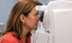
Retinal photography creates pictures of the back of the eye. These images can document findings and help your doctor compare changes between visits.
Some cameras capture a smaller area, while widefield systems photograph a larger portion of the retina. A wider image can be useful, but it does not guarantee that every important area is visible. Your doctor interprets photographs alongside your symptoms, medical history, and examination findings. The American Academy of Ophthalmology’s overview of fundus photography explains how these images support clinical evaluation.
Optical Coherence Tomography
OCT provides a different view. Instead of a conventional photograph, it uses light to create cross-sectional images of retinal tissue.
These scans help doctors evaluate retinal layers and structural changes that a surface photograph may not reveal. The American Academy of Ophthalmology’s explanation of OCT describes how the test helps assess retinal thickness and eye disease.
If your appointment includes a “retinal scan,” ask which test the office means. Retinal photography and OCT answer different clinical questions.
Why Your Doctor May Still Recommend Dilation
A retinal image may not provide everything your doctor needs to evaluate your eyes. Dilation creates a larger opening through which the doctor can examine the retina, optic nerve, and other internal structures.
The National Eye Institute’s guide to dilated eye exams explains how this examination helps identify conditions that may develop before you notice symptoms.
Your doctor may recommend dilation because of:
- New symptoms or a change in vision.
- Diabetes or other health conditions that affect the eyes.
- A personal or family history of eye disease.
- A finding that needs closer examination.
- An image that does not show enough detail.
The outer edges of the retina can be especially important when checking for retinal tears. Even ultra-widefield photography has limitations. A study comparing ultra-widefield images with examination for retinal horseshoe tears found that imaging alone missed some tears detected on clinical examination.
That does not make imaging unhelpful. It means a reassuring photograph is not always enough to rule out a problem.
When Having Both Adds Value
Imaging and dilation may serve different purposes during the same visit. Your doctor might examine the retina directly, photograph a finding for future comparison, or order OCT to assess a specific area more closely.
For example, a photograph can document the appearance of the optic nerve, while other testing measures its structure or function. A complete glaucoma evaluation may involve several tests because no single result tells the whole story.
A useful question is: “What information will this test add to my examination?”
That conversation helps you understand why your doctor recommends one test, both approaches, or additional testing.
What to Expect and How to Plan Your Visit

If your eyes are dilated, near vision may become blurry, and bright light may feel uncomfortable. Effects can last a few to several hours, depending on the drops and your individual response. The American Academy of Ophthalmology’s guidance on dilating eyedrops explains why the duration varies.
Before your visit:
- Bring sunglasses for the trip home.
- Arrange transportation if you are unsure you will feel comfortable driving.
- Allow flexibility for reading, screen work, or other close-up tasks afterward.
- Tell the care team about previous reactions to eye drops.
Do not drive while your vision is blurry or glare makes driving unsafe. Barnet Dulaney Perkins Eye Center’s guidance on preparing for your appointment can help you plan ahead.
Costs and Insurance: Ask What Is Included
Your out-of-pocket cost depends on the examination, the reason for testing, and your insurance benefits. Do not assume that every retinal image is included in the exam fee or that imaging and dilation have the same coverage.
Ask the office which tests are recommended, whether there is a separate charge, and which insurance plan may apply. Confirm benefits with your insurer and request an estimate when possible.
Review the practice’s accepted insurance information, but remember that plan participation does not guarantee coverage for every service.
Symptoms That Need Immediate Care
New flashes of light, a sudden increase in floaters, a curtain-like shadow, or sudden vision loss need immediate attention. Do not wait for a routine appointment or rely on a previous normal retinal image.
The National Eye Institute’s retinal detachment guidance advises seeing an eye doctor or going to an emergency room right away if you experience these symptoms.
For urgent eye concerns during office hours, call Barnet Dulaney Perkins Eye Center at (602) 955-1000 for emergency eye care guidance. If an eye care office is unavailable, go to the nearest emergency room. Do not drive yourself if your vision is impaired.
Understand Which Eye Tests You Need
You do not need to choose between retinal imaging and dilation on your own. At Barnet Dulaney Perkins Eye Center, your doctor can explain which tests your eyes need and why. Let your care team know if you have concerns about driving, returning to work, or the effects of dilating drops so you can plan your visit.
For a routine evaluation, schedule a comprehensive eye exam in Arizona and get clear answers about your examination options.