Keratoconus is a progressive condition where the front of the eye gradually thins and bulges outward, forming a cone-like shape.
The Role of the Cornea
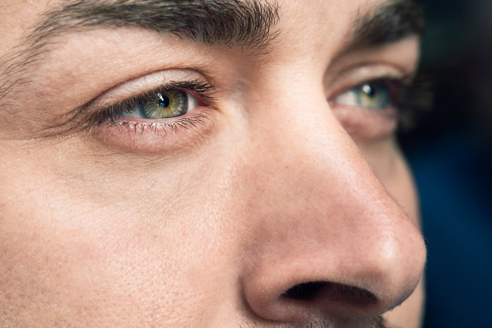
Understanding keratoconus helps us know how the cornea functions. The cornea is the clear, dome-shaped surface at the front of the eye. Normally round like a ball, it focuses light as it enters the eye. Light travels through the cornea, then through the pupil and lens, and finally reaches the retina at the back of the eye.
The shape of the cornea determines how accurately it directs light to the retina. When the cornea isn’t perfectly round—as in nearsightedness, farsightedness, or astigmatism—light may focus in front of or behind the retina, causing blurry vision.
What Happens in Keratoconus?
In keratoconus, the cornea thins and bulges forward into a cone shape. This altered shape distorts how light bends (refracts) as it enters the eye, resulting in blurred or warped vision.
Keratoconus is relatively rare. According to the Global Keratoconus Foundation, it affects 50 to 230 people per 100,000. In contrast, nearsightedness affects nearly 30% of the U.S. population. Keratoconus affects both men and women of all races and appears more frequently in individuals with Down syndrome. It usually begins during the teenage years.
Symptoms of Keratoconus
People developing keratoconus often notice persistent blurry vision, even after getting new prescriptions. They typically experience increasing nearsightedness and need frequent prescription updates. Other symptoms may include:
-
Sensitivity to light
-
Eye strain or general eye discomfort
-
Glare and trouble seeing at night, especially while driving
-
Headaches and eye irritation
Since other eye conditions can cause similar symptoms, seeing an eye doctor is essential. A proper diagnosis depends on both symptoms and direct corneal observation.
How Eye Doctors Diagnose Keratoconus
Eye doctors use a slit-lamp—a narrow beam of focused light combined with magnification—to examine the eye and detect corneal thinning or cone-shaped bulging. They also use corneal topography to measure the curvature of the cornea and assess how it reflects light.
9 Treatment Options for Keratoconus
Doctors select treatment based on the condition’s severity. Common options include:
1. Eyeglasses
In the early stages, simply updating your glasses prescription may be enough. Your doctor will continue to monitor your eyes for further changes.
2. Custom Soft Contact Lenses
Some manufacturers design soft contact lenses specifically for people with mild to moderate keratoconus.
3. Rigid Gas-Permeable Lenses
These hard lenses help maintain the cornea’s shape and allow light to focus correctly. They may be less comfortable and more difficult to fit than soft lenses.
4. Piggyback Contact Lenses
This approach uses both a soft and a hard lens. The soft lens sits on the eye for comfort, while the rigid lens on top provides optical correction.
5. Hybrid Contact Lenses
These lenses have a rigid center stabilizing the cornea’s shape and a soft outer ring for added comfort.
6. Corneal Implants (Intacs)
Doctors can insert small, curved plastic segments into the cornea to help reshape it. This minimally invasive procedure involves a tiny incision that heals quickly.
7. Corneal Collagen Cross-Linking (CXL)
This treatment strengthens the cornea by increasing the number of collagen bonds. Doctors apply riboflavin (vitamin B2) eye drops and activate them using ultraviolet light, helping the cornea reinforce its internal structure.
8. Corneal Transplant
In about 10% to 20% of cases, the cornea becomes too damaged and requires replacement with donor tissue. Patients often still need glasses or contact lenses afterward to achieve clear vision.
9. DALK (Deep Anterior Lamellar Keratoplasty)
This transplant technique replaces only the front and middle layers of the cornea. Because the innermost layers remain intact, the eye tends to heal faster and has a lower risk of rejection.
What Causes Keratoconus?
Doctors haven’t identified a single cause for keratoconus, which varies widely. Some people experience only mild changes, while others progress significantly.
Several factors may contribute to its development, though researchers haven’t confirmed whether these are causes or risk enhancers:
- Genetics: Less than 10% of people with keratoconus have a family history, but a hereditary link may still play a role in some cases.
- Enzyme Imbalance and Oxidative Stress: Studies suggest that keratoconus may involve an imbalance in corneal enzymes. This could affect how the eye repairs itself from oxidative stress, such as damage from free radicals.
- Eye Rubbing and Contact Lens Irritation: Chronic eye rubbing or irritation from poorly fitting contact lenses may weaken the cornea over time, though these links haven’t been definitively proven.
- UV Light Exposure: Excessive exposure to ultraviolet rays may also increase the risk of keratoconus.
Discuss Keratoconus Treatment Options With Your Eye Doctor
Since keratoconus develops gradually and shares symptoms with other conditions, seeing a cornea specialist for regular eye exams is essential, especially if you notice any loss of visual clarity.
At Barnet Dulaney Perkins Eye Center, we’re committed to protecting your vision. If you have questions or concerns about keratoconus, schedule an eye exam with one of our experts today.